Skip to content

MiraQ™ Ultimate

MiraQ™ Vascular

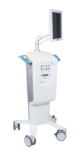
A dual technology for maximum precision and efficiency: The MiraQ™ system is the only one of its kind that combines ultrasound imaging and TTFM (transit time flow measurement). It provides instant feedback during procedures, vascular surgeons to make decisions and revisions in real time. This on-the-spot approach minimizes the risk of post-operative complications and additional surgeries, ensuring the best possible outcomes for patients.

MiraQ™ Ultimate is suitable for cardiac, vascular, and transplant surgery.

How MiraQ™ works
  • A high-frequency ultrasound imaging probe provides high-resolution near-field images. This technique, enhanced by color flow and Pulsed Wave (PW) Doppler modes, allows for the precise detection and visualization of blood flow and flow velocity.  
  • TTFM measures the difference in transit time of an ultrasound beam through blood flow. The TTFM probes utilize this technology to accurately measure blood volume flow intraoperatively. 
  • With the spatial information from ultrasound imaging and the quantitative data from TTFM, surgeons have real-time insights into the graft performance. 
  • The probes are approved for direct contact with the central circulatory system and heart (risk class III).  
Features & benefits
  • Documenting procedure: You can easily save flow tracings and ultrasound images on the device. The saved measurements can be used to create a PDF report on the surgical procedure. The report and saved measurements can be printed or saved on a USB stick. If DICOM is enabled, saved measurements and reports are accessible through PACS. 
  • Side-by-side screen assessments: You can compare any measurement against a reference measurement, evaluate improvement and perform functional tests on grafts, and store and report compared results with all values and indexes.
  • Easy OR integration: You can connect to external screens using display port and hospital information systems with DICOM.
  • Synchronized curves: You can interface other measurements (e.g. ECG or pressure) and have them synchronized with the flow curves for the most precise graft patency assessment. 
  • Convenient design: Interactive display, storage for manuals and cables, flexible monitor arm, rotatable screen. 
Fields of application
  • Cardiac surgery: The MiraQ™ system provides immediate feedback on the quality of the CABG surgery. Epicardial imaging shows coronary stenosis and graft anastomoses, while flow measurements verify graft patency. Epiaortic imaging may reveal aortic disease, which might prompt modifications of the surgical strategy.
  • Vascular surgery: The MiraQ™ system provides immediate feedback on the quality of peripheral bypass surgery, AV access surgery, and carotid endarterectomy. Flow and imaging in real time can secure graft patency and adequate flow levels. It may also reveal lumen defects which could cause postoperative stroke.
  • Transplant surgery: The MiraQ™ system ensures adequate hemodynamics and patent anastomoses during the procedure. Imperfect perfusion of the graft can be identified and instantly corrected to avoid primary graft dysfunction or graft infarction. 
Publications
Clinical retrospective study 2020

... results suggest that intraoperative PF [portal flow] plays an important role in determining early graft outcomes after DDLT [deceased donor liver transplantation].”  

Matsushima et al. Transplantation 2020. DOI: 10.1097/TP.0000000000002968

Observational cohort study 2019

“Intraoperative measurement of blood flow after reperfusion appears to be a predictor of EAD [early allograft dysfunction]; moreover, HAF [hepatic arterial flow] should be considered a predictor of 30-d patient mortality.”

Lominchar et al. World J Hepatol 2019. DOI: 10.4254/wjh.v11.i9.689