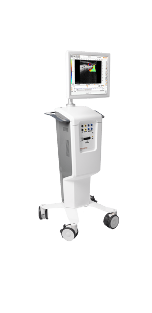
MiraQ™ Vascular

A dual technology for maximum precision and efficiency: The MiraQ™ system is the only one of its kind that combines ultrasound imaging and TTFM (transit time flow measurement). It provides instant feedback during procedures, allowing vascular surgeons to make decisions and revisions in real time. This on-the-spot approach minimizes the risk of post-operative complications and additional surgeries, ensuring the best possible outcomes for patients.
- A high-frequency ultrasound imaging probe provides high-resolution near-field images. This technique, enhanced by color flow and Pulsed Wave (PW) Doppler modes, allows for the precise detection and visualization of blood flow and flow velocity.
- TTFM measures the difference in transit time of an ultrasound beam through blood flow. The TTFM probes utilize this technology to accurately measure blood volume flow intraoperatively.
- With the spatial information from ultrasound imaging and the quantitative data from TTFM, surgeons have real-time insights into the graft performance.
- MiraQ™ Vascular identifies potential obstructions such as thrombus and restenosis, which can then be counteracted immediately.
- The probes are approved for direct contact with the central circulatory system and heart (risk class III).
- Documenting procedure: You can easily save flow tracings and ultrasound images on the device. The saved measurements can be used to create a PDF report on the surgical procedure. The report and saved measurements can be printed or saved on a USB stick. If DICOM is enabled, saved measurements and reports are accessible through PACS.
- Side-by-side screen assessments: You can compare any measurement against a reference measurement, evaluate improvement and perform functional tests on grafts, and store and report compared results with all values and indexes.
- Easy OR integration: You can connect to external screens using display port and hospital information systems with DICOM.
- Convenient design: Interactive display, storage for manuals and cables, flexible monitor arm, rotatable screen.
- peripheral bypass surgery
- carotid endarterectomy (CEA)
- hemodialysis through arteriovenous fistula (AV)
ESVS guidelines 2023
“For patients undergoing carotid endarterectomy, intra-operative completion imaging with angiography, duplex ultrasound or angioscopy should be considered in order to reduce the risk of peri-operative stroke.”
Naylor et al. Eur J Vasc Endovasc Surg 2023. DOI: 10.1016/j.ejvs.2022.04.011
Global vascular guidelines 2019
“Perform intraoperative imaging (angiography, DUS, or both) on completion of open bypass surgery for CLTI and correct significant technical defects if feasible during the index operation.”
Conte et al. J Vasc Surg 2019. DOI: 10.1016/j.jvs.2019.02.016
Prospective cohort study 2019
“The predictive value of intraoperative flow measurement is superior to intraoperative physical examination and could help reduce the fistula dysmaturation rate. Intraoperative transit time flow measurement is an easy method and can be used to predict successful fistula maturation in a high percentage rate.”
Meyer et al. J Vasc Access 2019. DOI: 10.1177/1129729819874312


